Bachelor program 2022-2023
Our bachelor program is an in-depth, extracurricular course for (bio-)medical students of the Radboud University.
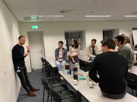
Below, you can find an impression of our bachelor program of 2022-2023. The topics that we discussed this year were: brain connections, gliomas, and tremors in Parkinson's disease.
Dr. Dylan Henssen
January 13th 2023
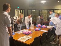
The evening started with an interactive lecture on neuroanatomy using 8 clinical cases. Then we went to the anatomical museum, where Dr. Henssen had arranged several preparations of real brains. In this way, the students could immediately apply the knowledge they had learned earlier in practice!
Dr. Mark ter Laan
January 20th 2023
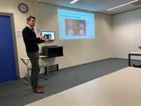
First, we were given an explanation of Broca and Wernicke's old-fashioned, topographical model and why this theory is outdated. In fact, brain functions appear to arise from networks and are not located in a universal location in the brain.
After the break, Dr. Ter Laan told us a lot about intra-operative techniques in the removal of gliomas, supported by visual material. Currently, there are many developments in this field, one even more interesting than the other.
Dr. Stephanie Forkel
January 27th 2023
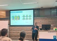
Dr. Forkel does a lot of research on the mechanism behind the production and processing of language and speech in the brain. During this lecture she explained to us, using her own research and state-of-the-art techniques, exactly how this works and what enables us to speak and interpret language.
She told us that the original model, where language and speech were assumed to be topographically organized by Broca and Wernicke, was too simplistic. This model mainly looked at the principle of localisationism of functions in the brain, while Dr. Forkel's new model looks at the associationism of functions in the brain instead, and thus of language and speech. Using these new insights, she has developed a new brain model of language and speech in the brain, with which the interindividual differences of speech and language can be represented. Using this model, the precise brain regions involved in language and speech can be looked at on an individual patient level in contrast to the population as a whole, and this can then, for example, be used in clinical practice to make predictions about the risk of aphasia in brain surgery or the predictive recovery capacity of brain functions after stroke.
Dr. Dylan Henssen
February 6th 2023
The lecture focused on the different types of imaging used within neuro-oncology: MRI, CT and PET. Dr. Henssen went over these techniques with us one by one, discussing the mechanism, clinical applicability and innovations of each technique.
Dr. Rick Helmich
February 13th 2023
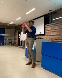
Using the latest research (from both Dr. Helmich himself and colleagues), we were taken through the pathological mechanism of tremor. We were taken through the latest theories and with which techniques research into these mechanisms is now being conducted.
We learned about different types of tremor and that each has its own mechanism. Dr. Helmich coupled this with the clinical presentation using visuals, which meant that this was a wonderful balance between clinic and neuroscience. In addition, we learned about different locations for deep brain stimulation for the different forms of tremor with their pros and cons and possible complications. This completed the circle of mechanism→clinic→therapy.
Finally, Dr. Helmich gave an example of a hugely interesting case study of a recent patient of his whose dystonic tremor completely disappeared after a CVA! With some imaging, footage and explanations, we were taken through this case. Naturally, there was plenty of room to ask questions during the lecture, which was enthusiastically taken advantage of!
This evening ended with an amazingly interesting discussion with the group, Dr. Helmich and of course some food and drinks. Here, everyone could ask all of their questions, both about the lecture and about Mr. Helmich's career and all the other things you wanted to know.
Craniotomy masterclass
February 7/8/9th 2023
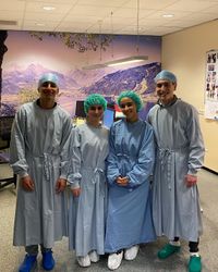
Last week we received lessons in small groups at the central animal laboratory. We were informed about the procedure regarding the request for animal research and that three bodies work closely together to regulate this. Then we saw pictures of some very interesting ex vivo models that have been used as research setups in recent years.
After this, we were given a further explanation of a craniotomy procedure in rats and we received suturing lessons. This was a tremendously interesting conclusion to a great undergraduate program!
